Advance Your Clinical Skills with One of the World’s Leading Educators
If you’re a physician, healthcare practitioner, biohacker, or wellness professional looking to expand your diagnostic and patient education skills, the Advanced Microscope Certification Program with Dr. Nick Delgado offers an exceptional opportunity.
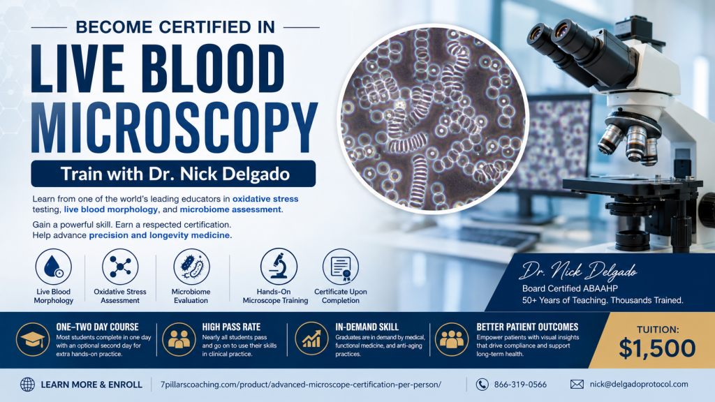
For over 50 years, Dr. Nick Delgado has trained thousands of physicians, scientists, nurses, and health professionals in live blood microscopy, hematology, oxidative stress assessment, and microbiome evaluation. As one of the world’s most recognized educators in anti-aging and lifestyle medicine, Dr. Delgado has helped practitioners integrate microscopy into patient education and wellness programs.
Rather than paying for repeated testing, learn to perform and interpret microscopy yourself.
Learn more:
https://7pillarscoaching.com/product/advanced-microscope-certification-per-person/
Certification Includes
- Live Blood Morphology
- Oxidative Stress Assessment
- Microbiome Evaluation
- Hands-on Phase Contrast Microscopy Training
- Certification Examination
- Certificate upon Successful Completion
Tuition: $1,500
Most students complete the course in one intensive day, with an optional second day available for additional hands-on practice before taking the certification examination. Nearly all students successfully complete the program.
Graduates have gone on to work in medical, functional medicine, regenerative medicine, and anti-aging practices. Several physicians currently rely on technicians trained by Dr. Delgado to evaluate every patient using microscopy as part of their educational process.
Precision Medicine Meets Functional and Lifestyle Medicine
Microscopy provides practitioners with a visual educational tool that helps patients better understand how nutrition, lifestyle, inflammation, and metabolic health affect the body.
Dr. Delgado teaches practitioners how to combine microscopy with standardized laboratory testing—including CLIA-waived Cholestech testing—to improve patient understanding and compliance.
Participants also receive guidance on selecting the appropriate microscope based on their practice goals and budget.
More information:
Why Live Blood Microscopy?
Nearly one million people die each year from chronic circulatory diseases that often develop silently over decades.
Modern microscopy allows practitioners to digitally capture and display live blood images, helping patients visualize aspects of their blood morphology while encouraging healthier lifestyle decisions.
Microscopy serves as an educational tool that complements traditional laboratory testing by illustrating textbook hematology principles in real time.
Practitioners learn how dietary choices, exercise, and nutritional interventions may influence blood appearance and metabolic health over time.
Visual Motivation Improves Patient Compliance
One of the greatest challenges in medicine is helping patients remain compliant with lifestyle recommendations.
Seeing one’s own blood under a microscope often creates a powerful educational experience.
For example, practitioners may observe changes in blood appearance following high-fat meals alongside fasting and postprandial laboratory testing.
Educational comparisons between:
- Glucose
- Triglycerides
- Lipid measurements
- Microscopic blood appearance
can help patients better understand how nutrition affects metabolic health.
As healthier lifestyle habits are adopted, repeat testing frequently demonstrates improvements in laboratory values and blood appearance, reinforcing positive behavioral changes.
Dry Blood Oxidative Stress Analysis
The certification also includes training in Dried Oxidative Blood Analysis, which evaluates dried blood samples for patterns associated with oxidative stress.
Participants learn to recognize various morphological configurations that have been proposed to correlate with oxidative damage and inflammatory processes.
Training discusses:
- Reactive Oxygen Toxic Species (ROTS)
- Free radical activity
- Oxidative stress
- Tissue inflammation
- Cellular degeneration
Healthy dried blood samples typically demonstrate consistent fibrin patterns and uniform coloration, while abnormal findings may suggest increased oxidative stress requiring further clinical evaluation.
Live Blood Morphology Training
Students learn how to observe and document:
- Red blood cell morphology
- White blood cell activity
- Lipid material
- Blood viscosity
- Signs of anemia
- Oxidative stress
- Inflammatory patterns
- Immune system activity
Training emphasizes microscopy as an educational adjunct used alongside appropriate clinical laboratory testing—not as a stand-alone diagnostic tool.
Understanding the Microbiome
The course also teaches one of the simplest visual methods for evaluating the oral microbiome.
Dental plaque collected from flossing is examined microscopically to demonstrate the balance between beneficial and potentially harmful bacteria.
Patients are often fascinated to observe bacterial diversity, including organisms such as spirochetes, making discussions about oral health, probiotics, nutrition, and lifestyle far more engaging.
Historical and Scientific Background
The course reviews historical and modern research relating to:
- Blood morphology
- Lipid metabolism
- Hematology
- Oxidative stress
- Diabetes
- Cardiovascular disease
- Nutrition
- Microbiology
- Prion research
Topics include discussion of the work of:
- Dr. Himsworth
- Dr. Ancel Keys
- Dr. Meyer Friedman
- Dr. Stanley Prusiner (1997 Nobel Prize in Physiology or Medicine)
- Additional investigators who have explored blood morphology, metabolism, and chronic disease.
Participants also review the evolving scientific literature concerning oxidative stress, inflammation, microbiome research, and lifestyle medicine.
Additional Educational Topics
The certification also discusses:
- Standardized laboratory testing
- Cholesterol metabolism
- Lipid peroxidation
- C-reactive protein
- Nitric oxide
- Insulin resistance
- Acid-base balance
- Reactive oxygen species
- Nutritional interventions
- Beta-glucans
- Superoxide Dismutase (SOD)
- Phytochemicals
- Lifestyle approaches to reducing oxidative stress
Learning Objectives
Participants will learn:
- How to perform live blood microscopy using phase contrast microscopy.
- How microscopy complements standardized laboratory testing.
- The relationship between blood viscosity, triglycerides, lipids, and chronic disease risk.
- Documentation and quantification techniques for inflammation, oxidation, anemia, infection, and additional wellness indicators.
- How to educate patients using visual microscopy findings to improve compliance.
Progress Monitoring
Students learn how to monitor patient progress through:
- Live blood microscopy (40x and 100x objectives)
- Dry blood microscopy (4x and 10x bright field)
- Oxidative stress markers
- Lipid profiles
- Cholesterol fractions
- Glucose testing
- Free fatty acids
- Toxic metal screening
- Salivary pH
- Salivary cortisol
Podcast
Precision Medicine Meets Functional Medicine, Lifestyle Medicine, Regenerative Medicine and Anti-Aging Medicine
Hosted by Dr. Nick Delgado featuring Dr. Anil Bajnath.
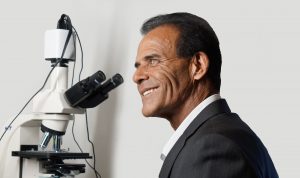
About Dr. Nick Delgado
Dr. Nick Delgado has trained and certified physicians in microscopy and hematology for over 50 years.
His mentors included:
- Phil Huckster, PhD
- Bob Bradford
- Ron Duvendack, MD
Dr. Delgado is the author of multiple books, including Annihilate Acne Now, and contributed to the Encyclopedia of Clinical Anti-Aging Medicine & Regenerative Biomedical Technologies.
He continues to compete internationally in strength endurance competitions and helped lead Team USA to victory in the World Championships.
Dr. Delgado is a Diplomate of the American Academy of Anti-Aging Health Practitioners (ABAAHP) and previously directed the Nathan Pritikin Better Health Program & Longevity Center.
Contact
Phone: 866-319-0566
Email: nick@delgadoprotocol.com
References
The educational program discusses published research including work by Swank, Scherer, Ginsberg, Lichtenstein, Kempner, West, Jenkins, Domingue, Kajander, McLaughlin, Nikkari, Tedeschi, Wintrobe’s Clinical Hematology, Stanley Prusiner, and numerous additional peer-reviewed publications listed in the original course syllabus.

